• Skip to main content
  • Skip to primary sidebar

EyeSurgeryClinics.org

eyesurgery

What Does 20/20 Vision Mean?

May 6, 2022 by eyesurgery

We all recognize the term “20/20 vision.” But what does 20/20 vision mean exactly? That’s what we will get into in this blog.

Plus, we will dive into what other kinds of visions there are and what they mean. Also, we’ll help you understand visual acuity and how doctors use the Snellen eye chart to diagnose eyesight conditions.

Eyesurgeryclinics.org provides several monthly blogs on eye-related topics. If you have an issue with your vision, you can read and learn more about it on our website. We aim to help those with sight problems identify and understand their condition.  We’re also an educational guide for aspiring optometrists and eye professionals.

20/20 Vision Definition

Firstly, what does 20/20 vision mean? Let’s start with the term visual acuity.

Visual acuity is the measure of how sharp your eyesight is. The standard eyesight test that determines your eyes’ sharpness takes place at a distance of twenty feet. The term 20/20 vision is what optometrists use to define normal vision. If you can see items and objects clearly from twenty feet away, doctors will deem you have 20/20 vision.

Visual acuity tests can be thorough, and they don’t always only test your forward vision. They may extend to your coordination, peripheral vision, focus, and depth perception. Doctors will have you look at various size letters from different angles to gauge your vision level.

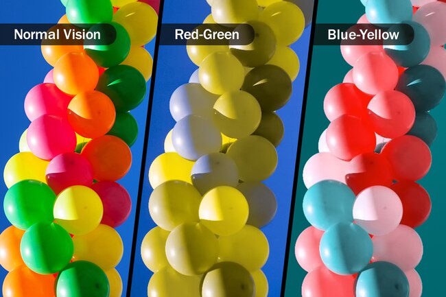
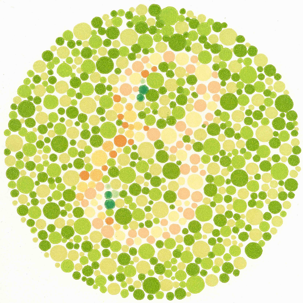
Even with in-depth testing, it can be hard to predict some eyesight issues you may have. For example, you may have trouble differentiating between colors and reading text with monochromatic backgrounds despite having 20/20 vision.

There are a few things that make for healthy visual acuity. Your cornea and lens’s job is to reflect light onto your retina. The efficiency of the lens and cornea dictates how sharp your vision is. Nerve sensitivity is also a factor in your visual acuity. Your nerves help protect your eye and allow you to process most visuals instantly.

People with clear eyesight do not require prescription glasses. However, as you get older, your sight may become worse. Just because you have 20/20 vision today doesn’t mean it will remain that way for the rest of your life.

You need to prove you have 20/20 vision to work some jobs. For example, 20/20 vision is the minimum eye strength that pilots need to fly a plane.

The Snellen Eye Chart

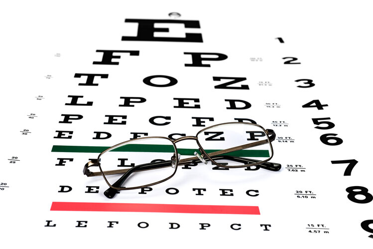
Though you may not recognize the name, most everyone knows what the Snellen eye chart is. Typically, your doctor or optometrist will place the Snellen chart in front of you and have you cover one eye. The chart has letters of all sizes, with the top row being the largest and the bottom row the smallest. You read the letters aloud, row by row.

This test is how doctors evaluate the sharpness of your vision. If you reach the eighth row of letters, your optometrist will determine that you have 20/20 vision. Doctors use this chart to diagnose a few different eye conditions. If you fail the test, you may require prescription glasses.

20/20 Variations

There are many different types of vision aside from 20/20. Firstly, there’s 20/15 eyesight. This level is better than its 20/20 counterpart. A person with 20/15 vision can see an item at twenty feet, what a person with 20/20 vision can only see standing fifteen feet away. Thus, people with 20/15 vision have better eyesight than those with 20/20.

Alternatively, there is 20/40 vision. Most everyone has at least this level of sight. It is the minimum eyesight you need to obtain a driver’s license in all fifty states. If you have lower than 20/40 vision, you will have difficulty reading books, paperwork, or any printed material.

Further down the line, we have 20/80 vision. An item on this level of eyesight is newspaper headlines. While not legally blind at 20/80, optometrists consider people with 20/80 moderately visually impaired, and you would require prescription glasses.

In some cases, you can correct 20/80 vision with eye surgery. Consult your optometrist about possible corrective procedures. Eyeglass technology has come a long way and is better than ever, and corrective eye surgery is becoming more common for those looking for a more permanent solution.

Finally, there is 20/200 vision. Optometrists will require you to wear eyewear at this level of sight. Plus, they will consider you legally blind. You need to stand twenty feet away from what people with 20/20 vision can see from 200 feet. However, people with 20/200 vision can still live a high-quality life.

Is 20/20 Vision Perfect?

Contrary to popular belief, 20/20 vision does not mean that you have perfect sight. Optometrists consider people with 20/20 vision to have normal eyesight.

It is possible to have more than 20/20 vision. People with this level of visual prowess may have some flaws in their peripheral vision or coordination. Your optometrist will let you know if your eyes need further evaluation after your Snellen test.

Many people who want to improve their eyesight turn to LASIK, a laser correction procedure. Ask your ophthalmologist about laser treatment to see if it is right for you. If it is, your ophthalmologist can refer you to a LASIK surgeon.

There are also eye specialists for athletes who feel their vision when playing sports is weak. Your optometrist can refer you to a sports specialist who can better diagnose your condition.

Read More

Most people have 20/20 vision. However, it is not a problem if you do not. As we get older, our eyesight diminishes, which is a part of life. The crucial thing is not to let it impact your day-to-day routine. With the proper treatment, you can find a way to cope with your eye disorder.

If you enjoy this article, feel free to browse more of our informational guides about other eye-related topics. We strive to help you learn all there is to know about eye conditions and terminology. Each year, we help thousands of people learn more about their eyesight issues and lead them on the path toward treatment.

Filed Under: Vision

Three Signs You Might Be Losing Your Sight

April 2, 2022 by eyesurgery

If you think you are losing your sight, read these three signs. If you are experiencing these symptoms, go to your ophthalmologist.  

headache

Severe and sudden eye pain. Possible vision problems can lead to sharp and sudden eye pain. If your optic nerve becomes inflamed, it will cause eye pain and vision loss. It can also lead to recurrent pain around or inside the eye. Ignoring the pain can lead to more serious vision damage. Pain in the eye will result in constant excruciating discomfort in the area. Eye pain that happens on the surface may cause itching, scratching, or burning sensation. Eye pain can be caused by a foreign object, infection, or trauma. Eye pain, which can be followed by vision loss, could be a symptom of an emergency medical issue. Schedule an appointment with your ophthalmologist as quickly as possible if you begin to lose your vision while experiencing eye pain.    

Hallucinations. We have made a list of common hallucinations that most people see when they have vision problems. Seeing flashes of lights and bright floating spots. When your vitreous gel inside your eye rubs against or pulls on your Renita, people say they saw flashing lights or light streaks. You may have experienced this odd sensation if you have ever had trauma to the eye and started to see “stars”. These flashes of lights will go on and off in several weeks or months. Floating “spider webs”. Most eye floaters are caused by age-related matters as the vitreous in your eye starts to become more liquid. Fibers within the vitreous tend to clump and can cast tiny shadows on your retina. The shadows you see are called floaters“curtain covering” one eye. A shadow or a “curtain” is described when vision is blocked by dark or grey shapes moving across the visual field. People have hallucinations when they lose their vision because their brain isn’t getting as much information as it was before. The brain responds by filling in the gaps with fantasy patterns or images stored in their brain. These stored images are what cause hallucinations.   

Other symptoms. Here are some more symptoms that could lead to vision loss. Sensing a “cup filling up with ink” in one eye, unusual, even painful, sensitivity to lights or glare. Swollen red eyes. Tiny blood vessels, which are invisible to the naked eye, can become red and swollen because of possible eye problems. Changes in the color of the iris. If one eye changes its color, it could be referred to as heterochromia. Changes in eye color can signal certain diseases. Any sudden change in vision. Any sudden change in vision could lead to vision loss. Ways this can happen are eye rubbing, dry eyes, crying, aging, or bad eyeglass prescription. White areas in the pupil. White spots on the eye can vary in how serious they are. Some can be not noticeable and others can cause lots of discomfort. Itching, burning, or a heavy discharge in the eye. A cause of combined eye burning, itching, and discharge in the eye, could possibly be an infection. Causes of the infection can include the herpes simplex virus, which causes cold sores and can also be spread to the eye.   

If you have these symptoms, call your ophthalmologist immediately to schedule an appointment, to make sure you don’t damage your eyes anymore. Always check out your eyes if you are feeling discomfort.  While waiting for your appointment, get a small towel wet with cold water and put it over your eyes. This will loosen your eyes until you get them checked out.  

Filed Under: Vision

Color Blindness

February 6, 2022 by eyesurgery

What is it? 

Color blindness or also known as color vision deficiency is a disorder in the eyes where the retina is not able to comprehend greens and reds correctly. Eyes use cones and rods to perceive color well, and if one of those cones is absent then you aren’t able to fully and accurately perceive light and color well. Rods detect are very sensitive to low light and can sense light and dark. Cones however are able to detect color when some are missing or one doesn’t work. If only one of the three cones doesn’t work properly, you have mild color blindness but if all three are absent then your color blindness is incredibly severe. 

COLOR BLINDNESS

Symptoms? 

Now although the symptoms seem pretty obvious seeing as the disorder is called color blindness, there is actually a bit of symptom and other disorders that this blindness could be linked to. For instance, color blindness can be associated with amblyopia, or better known as a lazy eye. Color blindness can affect the sharpness of vision when at its most severe state, and can lead to poor or worsening vision. Sensitivity to light is also found in this condition as well as a condition known as Nystagmus where the eyes move and shift randomly and rapidly. 

How do you get it? 

Color blindness is usually passed down from generation to generation, for example, if a mother has color blindness her son would probably be diagnosed with it as well. This is known as a congenital condition. If someone were to be diagnosed with it later in their life with no history of it in their family, it would most likely be from one of these causes. 

  • Toxic Buildup from drug usage 
  • Metabolic or Vascular disease 
  • Ordinary diseases 
  • Or Trauma 

Strangely, the color blindness received from diseases works differently from congenital color blindness. While when it is received congenitally, both eyes are affected more or less the same, but if it is contracted through disease then it can affect both eyes differently. If it is contracted congenitally, then the disorder will most likely keep its severity for its lifetime. Although, if it is contracted through a disease then the condition could worsen over time.  

Who is more at risk? 

 Men are usually at a bigger risk than women when it comes to being diagnosed with color blindness. In fact, a single man in every ten will have color blindness and most of those men come from Northern European descent. There are also conditions that may put people at risk for color blindness. These include,  

  • Glaucoma 
  • Diabetes 
  • Macular Degeneration 
  • Alzheimer’s  
  • Parkinson’s 
  • Chronic Alcoholism 
  • Leukemia 
  • Sickle Cell Anemia. 

There are also drugs that can encourage the diagnose of color blindness including Plaquenil. Plaquenil, which is also known as hydroxychloroquine helps with rheumatoid arthritis and other similar conditions.  

Treatment? 

A popular way to test someone for color blindness is a simple test that your ophthalmologist can conduct which includes finding a number hidden in vibrant dots. 

They are able to diagnose someone based on their ability to read and discover the number hidden inside. Now although it is fairly easy to find if someone has congenital color blindness, it is difficult to find how to treat them. Although we can’t necessarily fix it, there are special contact lenses and glasses that can fix the problems in the eyes. 

One of the tests here: 

Color Blind Test


How Is Color Blindness Tested? 

As written above, an easy and reliable way to test yourself is by using color “plates”. These plates are known as the Ishihara method and it is incredibly easy to test yourself. The only activity needed is to discover the number embedded into the shapes. There is no need to worry if you can’t decipher the plates though as according to Jane Edmond MD “it very minimally impacts everyday life”. Jane C. Edmond is an ophthalmologist at the Texas Children’s Hospital. Usually, color blindness doesn’t actually mean that someone cannot see colors. They can actually see colors quite clearly, but just don’t see them correctly. The most common kind of color blindness is red-green where the eyes can’t perceive these two correctly. This kind of blindness affects from 5% and up to 8% of males and only a small 0.5 % in females.  

Conclusion 

Although color blindness can be severe, it is usually solely congenital and can be easily helped with some special prescribed lenses. Even if color blindness cannot be remedied, it can be fixed by a trusted ophthalmologist. 

Filed Under: Vision

What Is Macular Degeneration: Symptoms, Diagnoses, and Treatments

January 16, 2022 by eyesurgery

If you or a loved one has been diagnosed with or might be suffering from macular degeneration, it is essential to stay informed and prepared.

Although age-related macular degeneration (AMD) is the leading cause of permanent vision loss in people aged 60 and above, receiving a diagnosis can be stressful and intimidating. You might be asking, “What is macular degeneration, and what are my treatment options?” Keep reading for information about the types, causes, symptoms, and treatment of age-related macular degeneration that will help you and your loved ones make the right choices for treatment and care.

What Is Macular Degeneration?

Age-related macular degeneration is a chronic, incurable disease of the eye. AMD directly impacts the macula—the central portion of the eye’s retina that contributes to our ability to focus our vision to see fine details, read, drive a car, etc. With AMD, the macula wears down or becomes damaged over time, creating central vision loss.

This type of vision loss most often impacts people over 60 and can worsen with time. Vision loss occurs in the central portion of a person’s field of sight, with peripheral and side vision typically remaining, though this vision is sometimes blurred or distorted.

Types of Macular Degeneration

Age-related macular degeneration presents as one of two main types: wet form and dry form.

Dry macular degeneration is the most frequently occurring form of age-related macular generation, comprising more than 80% of AMD diagnoses. With dry macular degeneration, the macula thins over time, and small protein deposits called drusen begin to form. Light sensitivity in the eye progressively weakens, leading to blind spots in or complete loss of one’s central vision.

Wet macular degeneration is significantly less common than dry macular degeneration and progresses differently. Wet macular degeneration occurs when abnormal, new blood vessels begin to form beneath the eye’s retina. When fluid or blood leaks from these blood vessels, vision can become distorted or appear “wavy.” Over time, fluid leakage in the retina causes scarring on the macula and can lead to blind spots in or loss of central vision.

There is another rare type of macular degeneration caused by a disease called Stargardt disease. Referred to as juvenile macular degeneration, this type of macular degeneration is not related to aging. Stargardt disease occurs in young people and is related to a recessive gene.

Macular Degeneration Causes and Risk Factors

Though research is ongoing, the exact cause or causes of age-related macular degeneration, wet or dry, are unknown. Scientists have determined that a combination of hereditary and lifestyle factors may trigger deterioration in the macula but have not yet determined how these factors contribute to macular degeneration.

Several risk factors potentially contribute to age-related macular degeneration, including:

  • Smoking
  • High blood pressure
  • Obesity
  • A diet high in saturated fat
  • Family history of age-related macular degeneration
  • Caucasian ancestry
  • Heart disease

Though the above are potential contributing factors, age is still the most significant risk factor for age-related macular degeneration. It is essential to regularly visit your ophthalmologist, who can give preventative recommendations and monitor for signs of macular degeneration.

Symptoms

Age-related macular degeneration may not be noticeable in its early stages. Vision distortion or loss may not become apparent until it is significant or is impacting both eyes. As the disease progresses, symptoms could include:

  • Unfocused or blurry vision, including difficulty reading or driving
  • Blind, blurred, or dark spots in the center of your vision

Changes in color perception are rare but possible. Age-related macular degeneration typically progresses in stages, throughout which symptoms may progress, relapse, or continually worsen.

Stages of Macular Degeneration

Early age-related macular degeneration is typically symptom-free and unnoticeable, outside of those subtle symptoms that a trained ophthalmologist can detect.

Intermediate age-related macular degeneration may present with some blurred or lost central vision but could also be symptom-free. An ophthalmologist can detect protein deposits and color changes in the retina that would indicate age-related macular degeneration is occurring.

Late age-related macular degeneration is when most vision loss has become noticeable and impacts day-to-day activities.

Early detection makes treatment options available that might help slow the progression of the disease. Regular visits to your ophthalmologist increase your chances of early detection, giving you your best probability of prolonging your vision.

Prevention

Following a healthy lifestyle is encouraged if you want to reduce your chances of developing age-related macular degeneration. Basic guidelines include:

  • Eating a diet rich in leafy greens
  • Wearing sunglasses with UV protection
  • Avoiding smoking
  • Exercising regularly

Though these efforts may reduce your chances of developing age-related macular degeneration, the best prevention is routine eye examinations. Tell your doctor about any changes or abnormalities in your vision, even if those changes seem insignificant at the time. If you are age 60 or above, ask your ophthalmologist to include age-related macular degeneration testing in every eye exam.

Diagnosis

Routine eye exams performed by trained ophthalmologists are the standard detection method for age-related macular degeneration. Doctors look for drusen protein deposits and for the growth of abnormal blood vessels in the retina that is typical in age-related macular degeneration.

Early detection is critical for prompt treatment, so doctors recommend routine eye exams. You can even perform an at-home tests for visual distortions using the Amsler Grid, a straight-line framework with defined axes and a central point. When viewed correctly, distortions in the Amsler Grid may indicate symptoms of age-related macular degeneration.

Treatment

Currently, age-related macular degeneration is incurable. Research into its causes and potential treatments is ongoing, however, and mitigation of the symptoms of macular degeneration is possible. Treatments range from anti-angiogenesis drugs, which block the creation of new blood vessels in the retina, to laser therapy, which actively destroys abnormal blood vessels in the eye.

Early detection of age-related macular degeneration gives a significant advantage to the treatment of the accompanying symptoms. If you’re concerned that you may be affected by macular degeneration or any eye or vision impairment, see your preferred ophthalmologist as soon as possible.

Filed Under: Vision

LASIK Aftercare

January 11, 2022 by eyesurgery

First, What is Lasik? 

LASIK is a refractive correction surgery that stands for Laser In-Situ Keratomileusis. This surgery is meant to fix any problems in the eye, more precisely in the cornea. If the cornea is curved or shaped incorrectly, it could lead to refractive errors in the eyes such as hyperopia or myopia. (Farsightedness and nearsightedness) This surgery works to attempt to reshape the clear layer of the eye known as the cornea. This will help fix any problems of light not entering the eye and not focusing on the retina correctly.  


LASIK cost


What does post-Lasik recovery entail? 

Although Lasik has an incredibly high 95% success rate, it includes quite an important recovery that could potentially damage your eyes if not taken care of correctly. This recovery includes visits with your surgeon, recovery plans, consulting, and specific guidelines. Lasik surgery includes creating an incision to make a tiny flap on the cornea, which then will allow the eye to reshape itself. This part is pretty simple as the only action required is to simply not damage the eye or the cornea. The tiny flap will soon reseal itself without a need for any medical supervision or stitches. Although there isn’t really anything to do when it comes to healing the wound, it is crucial to the recovery process that you do not rub your eyes or risk an eye infection in any way. While the cornea is reshaping itself, the eye is at its most vulnerable state, and messing with it in any way could dislodge the flap or cause a serious infection through the flap. This is why it is so important to touch your eyes as little as possible and limit their contact with anything at all, as even if you are sure that whatever it touches is clean it could still lead to a physical injury. After around 1 to 2 days, it is ideal to contact and check-in with your doctor to make sure that there isn’t any harm being done to your eyes and that the healing process is going normally. 

Timeline of Recovery 

During the first 24 hours, it is crucial to recovery that you avoid eye strain as much as possible to avoid damage to your vulnerable eyes. The American Refractive Surgery Council suggests that it should be possible to resume your daily activities and resume your normal life after around 2 days of recovery. The FDA has also suggested that it would be ideal to try and take a few days off of work to not have to run into early disturbances such as glares and halos. It is normal to experience strange and foggy vision as the eyes adjust themselves. It is also common to feel blurry vision for a little longer as well as some burning and itching as the anesthesia begins to wear off, although U.S NLM (National Library of Medicine) does report that this is completely normal and should begin to fade away after a few short hours. Once again to represent the importance of it, it is incredibly important to avoid any rubbing or bumping the eye. Although most blurriness is diminished, it is known that it’ll take up to 6 months for any vision problems to completely clear.  

This timeline is the usual and healthy line of recovery for Lasik. 

First Six Hours After Lasik 

  • Keep your eyes closed as much as possible and avoid glare and eye strain. Discomfort may be experienced as the eyes adjust to their temporary vulnerability. Your doctor may prescribe a medication to help the pain and even eye drops to protect from infection and lubricate the eye. 

The First Day Of Recovery 

  • The first 24 hours post Lasik are best spent avoiding eye strain as much as possible. This includes avoiding:
  1. Reading 
  2. Video Games 
  3. Television 
  4. Screen Time Such As Smartphone Usage 
  5. And Computers 

It is important to avoid these to not risk any damage to the cornea as to properly heal it back to full health. You will most likely receive an eye shield to help prevent the strain of the eyes and can shower as soon as a day after the surgery. Although it is a possibility to take a shower so soon after the surgery, it is important to keep any products or harmful chemicals out of your eyes that could burn or strain them. 

One to Two Weeks Post Lasik 

  • You can begin to exercise around a week after the surgery although it is important to avoid the Sun if you can. Avoid any makeup or any sort of cream or lotion around the eye. Swimming in any bodies of water that may be contaminated is not advised as contaminated water is a very easy point of access for diseases to enter the eye.  

From now on, it is pretty straightforward when it comes to recovering for the next couple of months. Just make sure to avoid large amounts of strain and screen time, and make sure to avoid UV rays and direct sunlight. If there are significant refractive errors, keep in mind that it could take longer to heal. 

Filed Under: LASIK

  • « Go to Previous Page
  • Page 1
  • Page 2
  • Page 3
  • Page 4
  • Go to Next Page »

Primary Sidebar

Pages

  • Home
  • About
  • Contact
  • Privacy Policy
  • Medical Disclaimer

Recent Posts

  • How Long Does Cataract Surgery Take?
  • What Is Strabismus?
  • The Dangers Of Lasik Eye Surgery
  • What is Glaucoma?
  • FarSightedness

Archives

  • October 2022
  • September 2022
  • August 2022
  • July 2022
  • June 2022
  • May 2022
  • April 2022
  • February 2022
  • January 2022

Categories

  • Cataracts
  • Glaucoma
  • LASIK
  • Vision

Medical Disclaimer

All content and media on this website are created and published online for informational purposes only. It is not intended as a substitute for professional medical advice and should not be relied on as health or personal advice.

Copyright © 2025 · Metro Pro on Genesis Framework · WordPress · Log in